Research Projects
TECHNOLOGY DEVELOPMENT
High-speed metabolic imaging in human brain
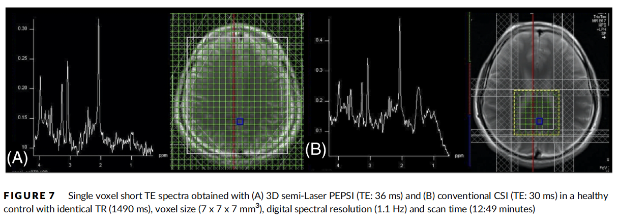
Proton magnetic resonance spectroscopic mapping (1H-MRSI) of brain metabolites can identify biomarkers relevant to psychiatric and neurological disease. There is currently increasing interest in extending 1H-MRSI techniques and processing capabilities to map J-coupled brain metabolite resonances. Glutamate (Glu) and glutamine (Gln) mapping are of particular interest because these metabolites are key components of energy metabolism and nitrogen homeostasis pathways and are also involved in excitatory synaptic neurotransmission. In vivo mapping of glutamate in clinically feasible acquisition times may have important diagnostic applications in psychiatric disorders and studies of aging. However, technical limitations, poor SNR, and data interpretation and analysis complications have prevented widespread use of MRSI in the clinical setting. Some of the most serious limitations of MRSI in the clinical setting are its intrinsically low SNR, long encoding times, limited volume coverage, limited spectral specificity and lack of absolute spectral quantification.
Proton-echo-planar-spectroscopic-imaging (PEPSI) developed in our laboratory has been successfully applied in a number of clinical applications, including panic disorder, autism spectrum disorder, and bipolar disorder. Our recent results using short echo time (TE) PEPSI in human brain demonstrate linear increase in sensitivity between 1.5 and 7 Tesla, and feasibility of 2-dimensional spatial mapping of J-coupled resonances at 3 and 4 T in less than 10 minutes.

We recently developed a novel approach for concurrent measurement of the tissue water and BOLD-contrast signal changes during MRSI acquisition by integrating spatial and spatial-spectral encoding modules into the water suppression module, that is, immediately after the water-excitation RF pulse and before application of the dephasing gradients. This approach enabled to demonstrate proof-of-concept for concurrent acquisition of task-based and resting-state fMRI with high spatial resolution, quantitative 3D PEPSI (fPEPSI).
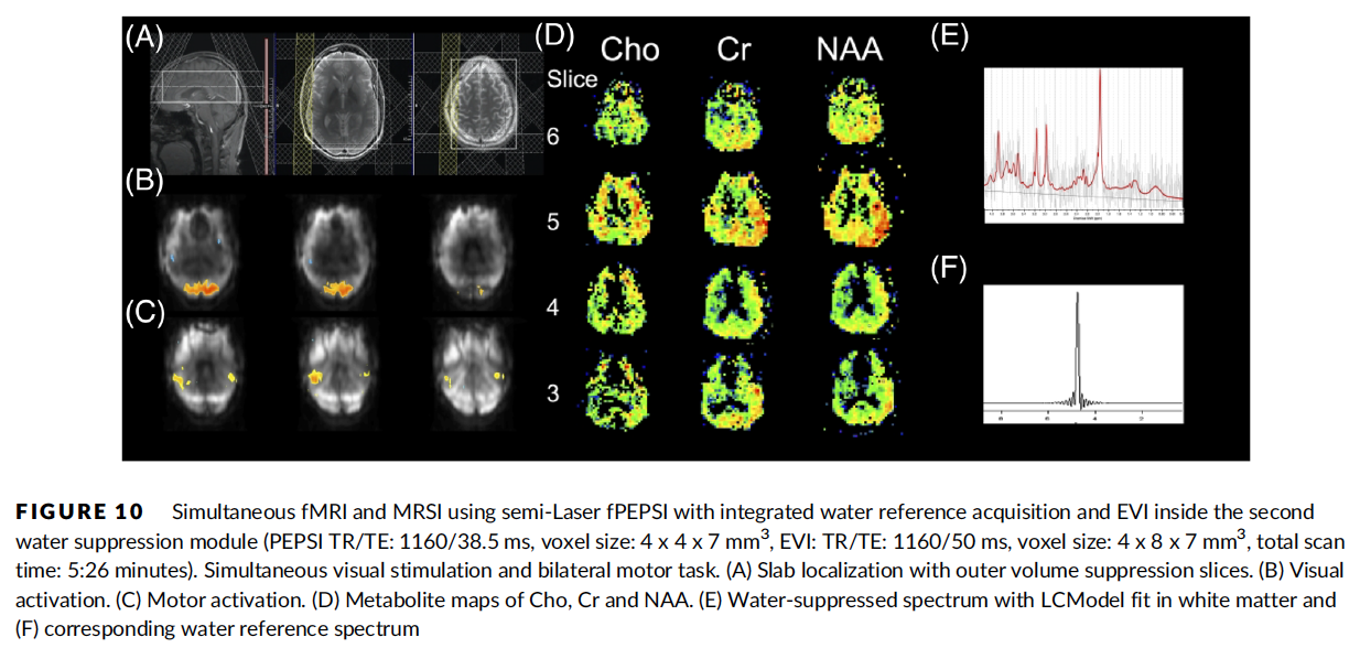
Ref: Posse et al. NMR Biomed. 2021
Measurement of brain metabolite diffusion has the potential to provide valuable information about intracellular changes associated with normal aging, in addition to diverse pathological processes, including ischemia, multiple sclerosis, brain tumors, and developmental disorders, such as autism spectrum disorder. The apparent diffusion coefficient (ADC) of metabolites reflects intracellular biophysical properties, including viscosity, cell swelling, restriction in subcellular structures, and cytoplasmic streaming. Metabolite fractional anisotropy (FA) provides additional directional information for characterizing the intracellular environment. The specificity of metabolite diffusion to the intracellular compartment provides unique information that cannot be obtained by diffusion tensor imaging (DTI), which measures water diffusion in both intra- and extracellular compartments.
Ref: Fotso et al., Magn Reson Med 2017
Real-time functional MRI
Functional MRI based on blood-oxygenation-level-dependent (BOLD) contrast is a technology that has found widespread application in cognitive neuroscience. At the same time, the sensitivity of data acquisition methodology has evolved to the point that brain activation can be detected in single trials and there is increasing interest in mapping brain activity in individual subjects for the purpose of understanding inter-individual differences in cognitive processing. Clinical applications for presurgical mapping and interactive brain-imaging-guided exams of patients suffering from psychiatric and neurological disorders are foreseeable.
Real-time fMRI is a variant of fMRI that enables monitoring of changes in brain activation during the ongoing scan. It is characterized by steady-state image reconstruction, preprocessing and statistical analysis in a time frame that is short with respect to the time to acquire a volume fMRI data set, and with a time delay from data acquisition that is shorter than the hemodynamic response delay, which is on the order of several seconds. Real-time fMRI offers new intriguing opportunities for monitoring brain processes related to thoughts and emotions. Using novel highly sensitive real-time data acquisition methods based on multi-echo Echo-Planar-Imaging (EPI) and real-time sliding-window correlation analysis, we have shown that it is possible to monitor dynamic changes in brain activation during brief motor, visual, auditory and cognitive tasks with an effective temporal resolution of just a few seconds. Recent real-time fMRI studies have demonstrated the feasibility of modulating brain activity in localized areas for the purpose of accelerated learning, to develop novel brain-computer interfaces for communication and for controlling pain perception in patients with chronic pain.
Our technology development is aimed at innovative individualized designs of fMRI experiments, which include, but are not limited to, (a) interactive brain-imaging-guided interview of patients suffering from psychiatric and neurological disorders that are refractory to conventional diagnosis and treatment, and (b) individualized training of mental abilities and control of brain activation patterns through the use of experimental feedback. The first approach is of importance in situations where the subject is either unable (e.g., stroke victims, babies and young children, many schizophrenic patients, many patients with major depression) or unwilling (e.g., in situations where deception is used) to accurately report his/her mental experience. The second approach is of interest for developing individually tailored training strategies for operators of complex machine-human interfaces (e.g., automobile driver, pilots) and for developing individually tailored mental learning strategies. Such capabilities would constitute a breakthrough in cognitive neuroscience, because they open the elusive world of human thought processes to rigorous neural systems level analysis.
VIDEO - Real-time fMRI of a Single Thought: Silent generation of a single word cued by a displayed letter. 50 s scan at 4 Tesla.

Refs: Mayer et al., Neuroimage; Vakamudi et al. Brain Connect 2020
Biophysics and Signal Physiology of functional MRI
The fast acquisition speed of multi-slab echo-volumar imaging enabled segregation of cardiac-related signal pulsation using ICA, which revealed distinct regional differences in pulsation amplitude and waveform, elevated signal pulsation in patients with arterio-venous malformations and a trend toward reduced pulsatility in gray matter of patients compared with healthy controls. Mapping cardiac pulsation in cortical gray matter may carry important functional information that distinguishes healthy from diseased tissue vasculature.
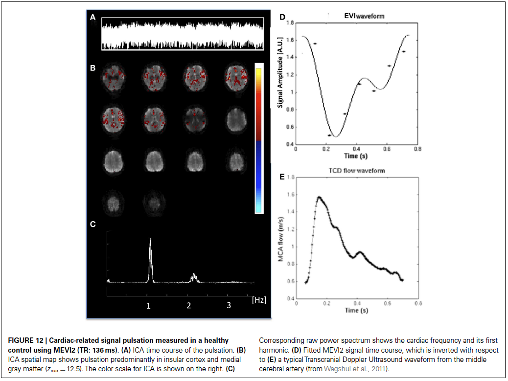
Refs: Posse et al., Front. Hum. Neurosci. 2013
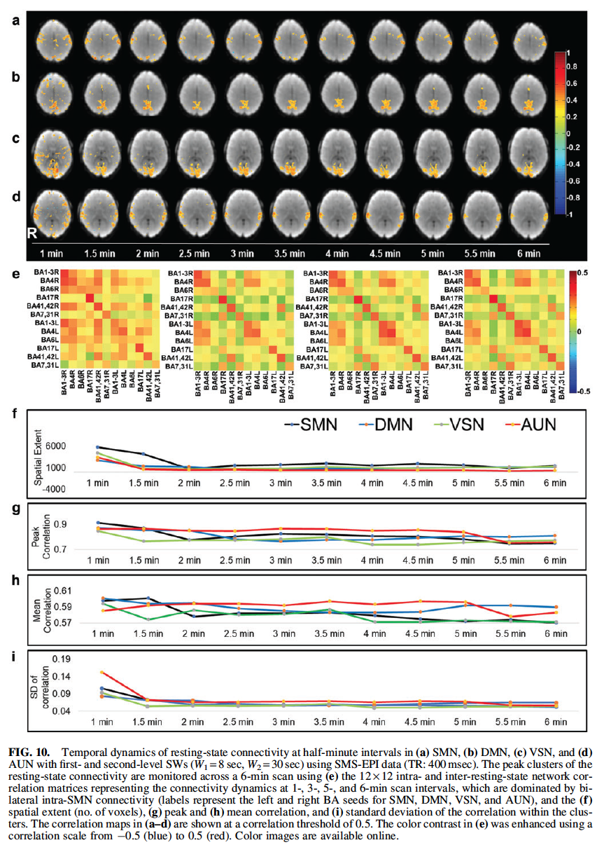
Current studies of resting-state connectivity rely on coherent signal fluctuations at frequencies below 0.1 Hz, however, recent studies using high-speed fMRI have shown that fluctuations above 0.5 Hz may exist. This study replicates the feasibility of measuring high frequency (HF) correlations in six healthy controls and a patient with a brain tumor while analyzing non-physiological signal sources via simulation. Resting-state data were acquired using a high-speed multi-slab echo-volumar imaging pulse sequence with 136 ms temporal resolution. Bandpass frequency filtering in combination with sliding window seed-based connectivity analysis using
running mean of the correlation maps was employed to map HF correlations up to 3.7 Hz. Computer simulations of Rician noise and the underlying point spread function were analyzed to estimate baseline spatial autocorrelation levels in four major networks (auditory, sensorimotor, visual, and default-mode).
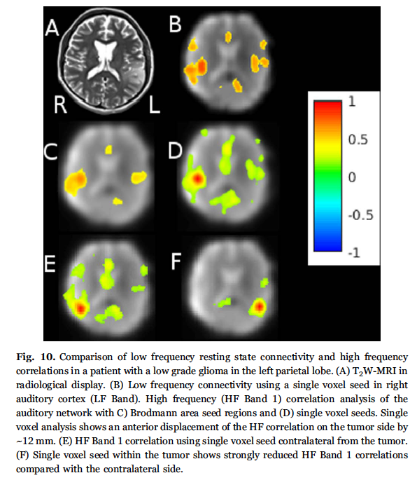
Ref: Trapp et al., Neuroimage 2018
Pattern classification in functional MRI
Pattern classification is a growing area of research in fMRI that aims at interpreting activation patterns in terms of underlying cognitive processes. Recent studies have demonstrated a high degree of pattern specificity for discrete neural processes and it is expected that pattern classification will play a major role in understanding cortical organization in individual subjects. Specifically, automatic interpretation and classification of neuroimaging data may hold important keys for understanding the human mind, which has raised interests due to the potential clinical applications. Information embedded in the spatial shape and extent of activation patterns, and differences in voxel-to-voxel time course, are not easily quantified with conventional analysis tools, such as statistical parametric mapping (SPM). Pattern classification in functional MRI (fMRI) is a novel approach, which promises to characterize subtle differences in activation patterns between different tasks. There is growing evidence that exquisite pattern specificity exists in visual cortex and other brain areas, such as motor cortex, auditory cortex and parietal cortex. However, automatic and reliable classification of patterns is challenging due to the high dimensionality of fMRI data, the small number of available data sets, inter-individual differences in activation patterns, and dependence on the image acquisition methodology. We recently introduced spatially distributed classifier for boosting to further reduce the dimensionality problem.
Our technology development is aimed at improving automated pattern classification to facilitate clinical applications of fMRI, such as fMRI-guided mental training, identification of disease markers (e.g. psychiatric disorders, epilepsy, migrane), and prediction of treatment response and relapse.
CLINICAL APPLICATIONS
Presurgical mapping in patients with brain tumors
Localization of eloquent cortex adjacent to brain lesions is of critical value in presurgical planning and decision-making. Mapping of resting-state networks (RSNs) using fMRI has been suggested as an alternative to task-based fMRI, however, the utility for presurgical planning is still under investigation. In this study, we characterize the sensitivity of novel high-speed resting-state fMRI for mapping eloquent cortex and disease-related changes in connectivity in patients with brain lesions.
The goal of this research is to develop a multi-modal imaging approach for presurgical mapping to guide neurosurgical resection and to minimize post-operative neurological deficits. Methods include task based and resting state ultra-high-speed real-time fMRI, 3D high-speed MR spectroscopic imaging, diffusion tensor MRI and concurrent fMRI-EEG, in correlation with a standard clinical workup that includes high-resolution MRI, MEG and PET.
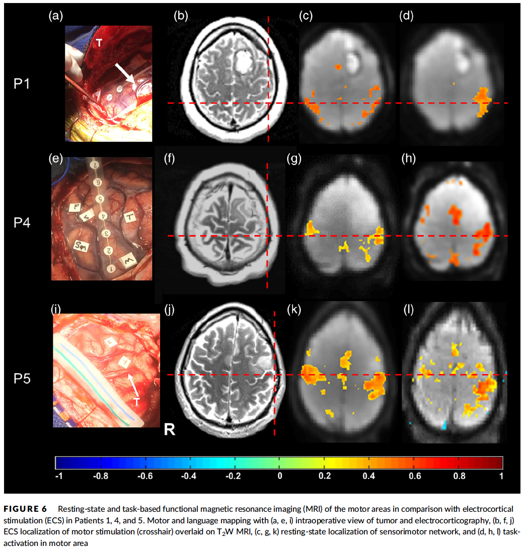
Refs: Vakamudi et al., Hum Brain Mapp. 2020; Posse et al., Front. Hum. Neurosci. 2013
High-speed metabolic imaging in brain tumors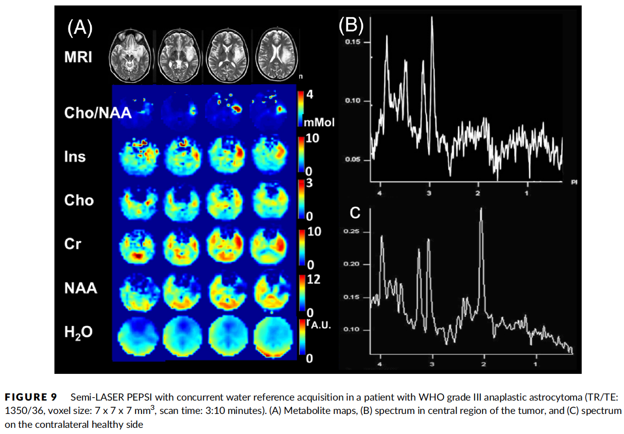
Ref: Posse et al. NMR Biomed. 2021
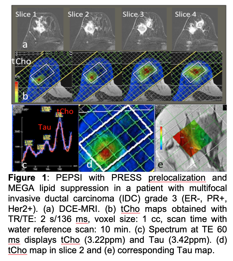
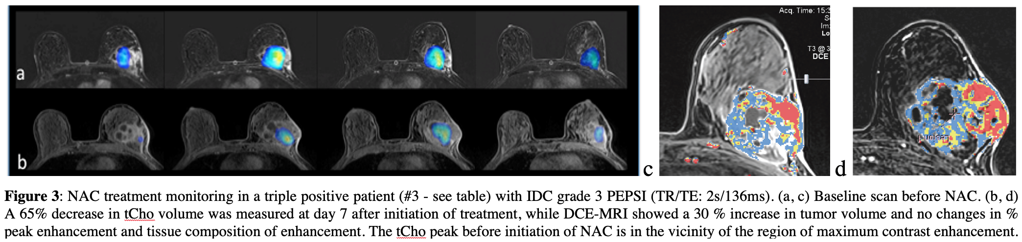
High-speed MR spectroscopic imaging of Choline in breast cancer
Magnetic resonance imaging (MRI) is playing an increasingly important role in clinical diagnostics of breast cancer, including screening for breast cancers in high risk women. However, overall specificity has been low, resulting in a considerable number of benign biopsies. Recent studies reported that adding quantitative MR spectroscopy (MRS) results (mostly focusing on total choline) to a dynamic contrast enhanced (DCE) MRI exam produced improvements in the sensitivity, specificity, and accuracy for all readers, and improved the inter observer agreement between the readers. A second promising application of breast MRS involves predicting response to treatment. However, the majority of breast MRS studies to date have used single-voxel spectroscopy (SVS) to localize the spectrum to a single volume centered on the lesion of interest, which does not allow characterization of lesion heterogeneity.
Our research aims at developing high-speed proton MR Spectroscopic Imaging (MRSI) methodology based on Proton-Echo-Planar-Spectroscopic-Imaging (PEPSI) to map total Choline to characterize lesion heterogeneity and improve the specificity of a Breast MR exam for disease staging and for treatment monitoring. Accurate early identification of treatment failure or success could save significant time and resources, and minimize patient risk and side effects in evaluation of any new therapy.